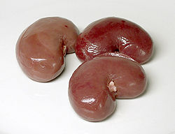
The kidneys are paired organs, which have the production of urine as their primary function. Kidneys are seen in many types of animals, including vertebrates and some invertebrates. They are part of the urinary system, but have several secondary functions concerned withhomeostatic functions. These include the regulation of electrolytes, acid-base balance, and blood pressure. In producing urine, the kidneys excrete wastes such as urea and ammonium; the kidneys also are responsible for the reabsorption of glucose and amino acids. Finally, the kidneys are important in the production of hormones including vitamin D, renin and erythropoietin.
Located behind the abdominal cavity in the retroperitoneum, the kidneys receive blood from the paired renal arteries, and drain into the pairedrenal veins. Each kidney excretes urine into a ureter, itself a paired structure that empties into the urinary bladder.
Renal physiology is the study of kidney function, while nephrology is the medical specialty concerned with diseases of the kidney. Diseases of the kidney are diverse, but individuals with kidney disease frequently display characteristic clinical features. Common clinical presentations include the nephritic and nephrotic syndromes, acute kidney failure, chronic kidney disease, urinary tract infection, nephrolithiasis, and urinary tract obstruction.[1]
Anatomy[edit]LocationIn humans, the kidneys are located behind the abdominal cavity, in a space called the retroperitoneum. There are two, one on each side of thespine; they are approximately at the vertebral level T12 to L3.[2] The right kidney sits just below the diaphragm and posterior to the liver, the left below the diaphragm and posterior to the spleen. Resting on top of each kidney is an adrenal gland (also called the suprarenal gland). The asymmetry within the abdominal cavity caused by the liver typically results in the right kidney being slightly lower than the left, and left kidney being located slightly more medial than the right.[3][4] The upper (cranial) parts of the kidneys are partially protected by the eleventh and twelfthribs, and each whole kidney and adrenal gland are surrounded by two layers of fat (the perirenal and pararenal fat) and the renal fascia. Each adult kidney weighs between 125 and 170 g in males and between 115 and 155 g in females.[2] The left kidney is typically slightly larger than the right.[citation needed]
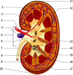
[edit]Structure1. Renal pyramid • 2. Interlobar artery • 3. Renal artery • 4. Renal vein • 5. Renal hilum • 6. Renal pelvis • 7. Ureter • 8. Minor calyx • 9. Renal capsule • 10. Inferior renal capsule • 11. Superior renal capsule • 12. Interlobar vein • 13. Nephron • 14. Minor calyx • 15. Major calyx • 16. Renal papilla• 17. Renal columnBean-shaped structures, each kidney has concave and convex surfaces. The concave surface, the renal hilum, is the point at which the renal artery enters the organ, and the renal vein andureter leaves. The kidney is surrounded by tough fibrous tissue, the renal capsule, which is itself surrounded by perinephric fat, renal fascia (of Gerota), and paranephric fat. The anterior (front) border of these tissues is the peritoneum, while the posterior (rear) border is thetransversalis fascia.
The substance, or parenchyma, of the kidney is divided into two major structures: superficial is the renal cortex and deep is the renal medulla. Grossly, these structures take the shape of 8 to 18 cone-shaped renal lobes, each containing renal cortex surrounding a portion of medulla called a renal pyramid (of Malphigi).[2] Between renal pyramids, which are composed of medulla, are projections of cortex called renal columns (of Bertin). Nephrons, the urine-producing functional structures of the kidney, span the cortex and medulla. The initial filtering portions of the nephron, the renal corpuscles, are located in the cortex and each sends a renal tubule that passes from the cortex deep into the medullary pyramids. Part of the renal cortex, amedullary ray is a collection of renal tubules that drain into a single collecting duct.
The tip, or papilla, of each pyramid empties urine into a minor calyx, minor calyces empty intomajor calyces, and major calyces empty into the renal pelvis
[edit]Blood supplyThe kidneys receive blood from the renal arteries, left and right, which branch directly from theabdominal aorta. Despite their relatively small size, the kidneys receive approximately 20% of the cardiac output.[2]
Each renal artery branches into segmental arteries, dividing further into interlobar arteries which penetrate the renal capsule and extend through the renal columns between the renal pyramids. The interlobar arteries then supply blood to the arcuate arteries that run through the boundary of the cortex and the medulla. Each arcuate artery supplies several interlobular arteries that feed into the afferent arterioles that supply theglomeruli.
After filtration occurs the blood moves through a small network of venules that converge into interlobular veins. As with the arteriole distribution the veins follow the same pattern, the interlobular provide blood to the arcuate veins then back to the interlobar veins which come to form the renal vein exiting the kidney for transfusion for blood.
